'%3e%3cpath%20d='M706.453%20375.999C860.234%20375.999%20984.899%20291.829%20984.899%20188C984.899%2084.1703%20860.234%200%20706.453%200C552.671%200%20428.006%2084.1703%20428.006%20188C428.006%20291.829%20552.671%20375.999%20706.453%20375.999Z'%20fill='%23009B4F'/%3e%3cpath%20d='M278.447%20375.999C432.229%20375.999%20556.893%20291.829%20556.893%20188C556.893%2084.1703%20432.229%200%20278.447%200C124.665%200%200%2084.1703%200%20188C0%20291.829%20124.665%20375.999%20278.447%20375.999Z'%20fill='%23009B4F'/%3e%3cpath%20d='M141.484%20166.68C125.01%20162.158%20114.996%20158.927%20114.996%20150.529C114.996%20143.422%20122.102%20138.9%20133.085%20138.9C144.068%20138.9%20155.051%20143.099%20164.095%20150.529L164.418%20150.852L165.388%20129.855C156.666%20125.01%20147.944%20121.78%20133.731%20121.78C123.394%20121.78%20114.027%20124.364%20107.243%20129.209C99.8137%20134.378%2095.9374%20142.13%2095.9374%20151.175C95.9374%20168.941%20110.796%20176.694%20123.394%20179.924C140.192%20184.446%20151.821%20188.323%20151.821%20198.659C151.821%20207.381%20144.068%20212.872%20131.47%20212.872C118.872%20212.872%20106.274%20207.381%2096.5834%20198.013L96.2604%20197.69L94.3223%20219.979C104.336%20226.762%20116.934%20230.316%20130.501%20230.316C142.13%20230.316%20152.144%20227.408%20159.25%20221.917C167.003%20216.103%20171.202%20207.704%20171.202%20197.367C171.202%20181.862%20161.511%20171.848%20141.484%20166.68Z'%20fill='white'/%3e%3cpath%20d='M264.557%20188C264.557%20215.78%20246.467%20230.316%20225.471%20230.316C215.457%20230.316%20207.381%20226.762%20201.244%20221.271V253.896H182.186V146.976H201.244V156.02C207.704%20149.883%20216.103%20145.684%20226.763%20145.684C247.436%20145.36%20264.557%20160.22%20264.557%20188ZM245.498%20188C245.498%20172.494%20236.131%20162.158%20222.887%20162.158C214.811%20162.158%20207.058%20165.711%20201.567%20173.787V203.182C207.058%20210.288%20214.488%20213.841%20222.887%20213.841C236.131%20213.841%20245.498%20203.505%20245.498%20188Z'%20fill='white'/%3e%3cpath%20d='M352.095%20194.137H294.274C296.535%20207.058%20305.903%20213.841%20318.824%20213.841C329.807%20213.841%20337.882%20211.257%20346.604%20206.089L347.573%20222.886C339.82%20227.085%20330.13%20230.316%20317.532%20230.316C294.274%20230.316%20274.893%20215.78%20274.893%20188.323C274.893%20162.804%20294.274%20145.683%20317.209%20145.683C341.112%20145.683%20353.71%20160.22%20353.71%20181.216C353.71%20185.415%20353.064%20189.615%20352.095%20194.137ZM335.621%20178.632C335.621%20168.618%20329.161%20160.866%20317.532%20160.866C306.549%20160.866%20296.858%20168.295%20294.597%20180.893H335.298C335.621%20180.247%20335.621%20179.601%20335.621%20178.632Z'%20fill='white'/%3e%3cpath%20d='M443.189%20221.917L445.128%20203.505C452.88%20209.965%20463.54%20213.841%20474.846%20213.841C482.921%20213.841%20488.413%20210.611%20488.413%20204.797C488.413%20189.615%20444.482%20197.367%20444.482%20169.91C444.482%20153.436%20458.695%20145.36%20475.492%20145.36C487.444%20145.36%20496.488%20148.268%20503.272%20152.467L502.303%20169.587C494.227%20164.096%20484.213%20161.189%20475.492%20161.189C468.708%20161.189%20463.217%20163.773%20463.217%20168.941C463.217%20181.862%20507.471%20175.402%20507.471%20203.828C507.471%20221.271%20492.289%20229.993%20474.523%20229.993C461.602%20230.316%20450.942%20227.085%20443.189%20221.917Z'%20fill='white'/%3e%3cpath%20d='M590.488%20177.986V228.701H571.429V220.302C564.969%20226.439%20556.57%20229.993%20546.88%20229.993C528.467%20229.993%20517.484%20219.01%20517.484%20203.505C517.484%20187.03%20530.405%20177.017%20549.464%20177.017C556.57%20177.017%20563.677%20178.309%20571.752%20180.57V177.986C571.752%20165.388%20562.385%20159.897%20550.756%20159.897C542.034%20159.897%20532.02%20163.127%20524.914%20168.941L524.268%20152.467C530.405%20148.268%20541.711%20145.037%20554.309%20145.037C574.983%20145.36%20590.488%20155.374%20590.488%20177.986ZM571.429%20206.735V194.46C564.646%20192.199%20558.185%20190.907%20551.402%20190.907C541.711%20190.907%20535.574%20196.075%20535.574%20203.505C535.574%20210.934%20542.034%20215.78%20550.433%20215.78C558.508%20215.78%20564.969%20212.872%20571.429%20206.735Z'%20fill='white'/%3e%3cpath%20d='M592.104%20146.976H612.777L636.358%20198.659L659.616%20146.976H680.289L641.526%20228.701H630.866L592.104%20146.976Z'%20fill='white'/%3e%3cpath%20d='M754.907%20194.137H697.409C699.67%20207.058%20709.038%20213.841%20721.959%20213.841C732.941%20213.841%20741.017%20211.257%20749.739%20206.089L750.708%20222.886C742.955%20227.085%20733.264%20230.316%20720.667%20230.316C697.409%20230.316%20678.027%20215.78%20678.027%20188.323C678.027%20162.804%20697.409%20145.683%20720.343%20145.683C744.247%20145.683%20756.845%20160.22%20756.845%20181.216C756.522%20185.415%20756.199%20189.615%20754.907%20194.137ZM738.756%20178.632C738.756%20168.618%20732.295%20160.866%20720.667%20160.866C709.684%20160.866%20699.993%20168.295%20697.732%20180.893H738.433C738.433%20180.247%20738.756%20179.601%20738.756%20178.632Z'%20fill='white'/%3e%3cpath%20d='M826.618%20148.914L824.034%20166.357C819.511%20163.773%20814.989%20163.127%20810.467%20163.127C802.068%20163.127%20794.315%20167.326%20788.824%20176.371V228.701H769.766V146.976H788.824V157.958C794.315%20150.529%20802.068%20145.36%20812.728%20145.36C817.896%20145.36%20822.418%20146.653%20826.618%20148.914Z'%20fill='white'/%3e%3cpath%20d='M832.109%20221.917L834.048%20203.505C841.8%20209.965%20852.46%20213.841%20863.766%20213.841C871.841%20213.841%20877.333%20210.611%20877.333%20204.797C877.333%20189.615%20833.401%20197.367%20833.401%20169.91C833.401%20153.436%20847.615%20145.36%20864.412%20145.36C876.364%20145.36%20885.408%20148.268%20892.192%20152.467L891.223%20169.587C883.147%20164.096%20873.133%20161.189%20864.412%20161.189C857.628%20161.189%20852.137%20163.773%20852.137%20168.941C852.137%20181.862%20896.391%20175.402%20896.391%20203.828C896.391%20221.271%20881.209%20229.993%20863.443%20229.993C850.522%20230.316%20839.862%20227.085%20832.109%20221.917Z'%20fill='white'/%3e%3cpath%20d='M429.298%20206.412C422.838%20211.257%20416.054%20213.841%20408.302%20213.841C395.058%20213.841%20383.429%20204.474%20383.429%20188C383.429%20171.525%20395.058%20162.158%20408.302%20162.158C416.054%20162.158%20422.838%20164.742%20429.298%20169.264C430.268%20163.45%20431.237%20157.958%20432.852%20152.467C426.068%20148.591%20417.67%20145.683%20406.041%20145.683C382.783%20145.683%20363.725%20161.512%20363.725%20188C363.725%20214.487%20382.783%20230.316%20406.041%20230.316C417.67%20230.316%20426.068%20227.085%20432.852%20222.886C431.237%20217.395%20429.944%20211.903%20429.298%20206.412Z'%20fill='white'/%3e%3c/g%3e%3cdefs%3e%3cclipPath%20id='clip0_1478_9029'%3e%3crect%20width='984.9'%20height='375.999'%20fill='white'/%3e%3c/clipPath%3e%3c/defs%3e%3c/svg%3e)
1 of 6
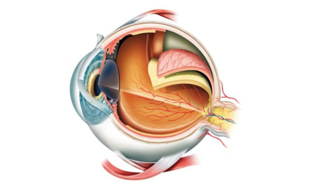
Cornea
This is a see‑through, dome‑shaped front layer that covers the iris, pupil, and anterior chamber. It provides about 70% of your eye's focusing power by refracting (bending) light as it enters. Irregularities in the cornea's shape can cause astigmatism, where light doesn't focus properly on the retina.
2 of 6
Iris and pupil
The iris is the coloured part of your eye containing muscles that control the pupil size. The pupil is the dark opening that allows light to enter. In bright conditions, the pupil constricts (gets narrower); in dim light, it dilates (gets wider) to let more light in.
3 of 6
Lens
Located behind the iris, the lens provides 30% of your eye's focusing power and can change shape for near and distance vision. As we age, the lens loses flexibility, leading to presbyopia where near vision becomes difficult.
4 of 6
Retina
The light‑sensitive tissue lining the back of your eye that converts light into electrical signals. The retina contains photoreceptors (rods and cones) that detect light and colour, sending signals via the optic nerve to your brain.
5 of 6
Aqueous Humour
The clear fluid filling the front chambers of your eye. It maintains eye pressure, provides nutrients to tissues without blood vessels, and removes waste. Problems with drainage can lead to glaucoma.
6 of 6
Vitreous Humour
The gel‑like substance filling the large cavity behind the lens. It helps maintain the eye's shape and allows light to pass through to the retina. Changes with age can cause floaters and flashes.
